Cells: Variation in Number, Shape, and Size
All living things carry out certain basic functions with the help of different sets of organs. Cells are called the structural unit of an organ. These may be compared to the bricks of a wall. As bricks are assembled to make a wall, similarly, cells are assembled to form an organism. Let us find out more about cells.
The smallest structural and functional unit of an organism is called a cell. Cells of organisms show variation in terms of their number, shape, and size. Let us discuss each of these variations in detail.
Variation in Number
All living organisms are made up of one or more cells. On the basis of cell number, organism are grouped into 2 categories. Bodies of organisms may consist of one or many cells. Organisms whose body consists of a single cell are called unicellular organisms. Examples of unicellular organisms are Amoeba, Paramoecium, Euglena, and bacteria. Thus, in a unicellular organism, a single cell performs all vital activities like feeding, movement, respiration, and reproduction. Organisms whose body consists of many cells are called multicellular organisms. For example – Plants, animals & fungi. Most plants and animals (including human beings) are multicellular organisms.
Variation in Size
Most cells are microscopic and cannot be seen with the naked eye. Cell size may vary from a micrometre (a millionth of a metre) to a few centimetres. The smallest cells are bacteria, which generally range in size from 0.1 to 0.5 micrometre. The largest cell is the egg of an ostrich, which is 170 millimetre in diameter. Human nerve cells are believed to be the longest cells.
- The size of different cells ranges between broad limits.
- Some plants and animals cells are visible to the naked eye.
- Most cells are visible only with microscope.
- The prokaryotic cells usually range between 1 to 10 mm.
- The eukaryotic cells usually range between 10 to 100 mm.
- Amoeba proteus may reach a diameter of 0.5 mm.
- The smallest cells are those of Mycoplasma laidlawiil (0.1µ in dimeter) or PPLO (pleuro pneumonia like organism).
- The largest cell is egg of an Ostrich.
Variation in Shape
Cells exist in different shapes. They can be disc-shaped, polygonal, rectangular, branched, or even irregular. The shape of a cell depends on the specific function it performs. Here are a few examples that illustrate this.
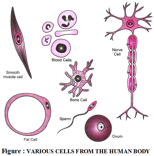
- The shape of cell may be variable or fixed.
- Variable shape occur in Amoeba, WBC etc.
- Fixed shape occur in most plant and animals.
- Cells may be diverse shapes such as polyhedral (8, 12 or 14 sides) spherical (e.g. eggs of mainly animals), spindle shaped (Smooth muscle fibres), elongated (e.g. Nerves cells) so on.
Nerve cells carry messages between different parts of the body. Hence, they are elongated in shape. Muscle cells help in movement through contraction and expansion. Hence, they are thin and long. Skin cells cover a large area. Hence, they are flat in shape.
Activity
Aim: To observe a single cell (hen’s egg).
Materials needed: A hen’s egg, a boiling pan, and water.
Method: Boil the egg and remove the shell. Cut the boiled egg into two halves.
Observation: The boiled egg has a yellow part and a white part surrounding it. The white part is called albumin while the yellow part is called yolk. In an unboiled egg, the albumin is a jelly-like transparent liquid. The yolk looks like a thicker yellow jelly.

PREPARING A SLIDE TO VIEW CELLS
Most cells are viewed with the help of a compound microscope. To view a specimen under a microscope, it has to be first placed Inanimate Not alive on a glass slide. Placing the specimen on the slide is called mounting. Two types of mounts are generally prepared in the laboratory: dry mount and wet mount.
A dry mount is generally used for viewing inanimate objects. As the term suggests, a dry mount does not require water. Wet mounts are generally prepared using water and are used for viewing living specimens like organisms and cells. In a wet mount, a small piece of the specimen is placed at the centre of the slide with one or two drops of water. The specimen is then covered with a coverslip and viewed under a microscope. In case of permanent slides (i.e., slides that need to be preserved for later use), materials other than water need to be used as the specimens have to be preserved for a longer duration.
The specimen to be viewed under a microscope is often stained (i.e., coloured) with a dye. Staining highlights biological tissues and specific regions in the cells, which makes it easier for us to view the details. Some of the commonly used staining dyes are safranin, methylene blue, and crystal violet.
Prokaryotic and Eukaryotic Cells
Cells that lack a well-defined nucleus surrounded by a nuclear membrane are called prokaryotic cells. Organisms that have such cells are called prokaryotes (pro, primitive; karyon, nucleus). Examples of prokaryotes are bacteria and blue-green algae. Cells that have a well-defined nucleus are called eukaryotic cells. Organisms that have such cells are called eukaryotes (eu, true; karyon, nucleus). All organisms apart from bacteria, blue-green algae, and viruses are eukaryotes.
Cell Division and Growth
The food that we eat leads to an increase in the size of cells. After reaching a certain size, a cell divides into two by a process known as cell division. Increase in the number of cells in the body as a result of cell division is responsible for growth in organisms. Cell division also replaces the dead or damaged cells with new ones and is, thus, responsible for healing wounds.