Selina Concise Biology Class 10 ICSE Solutions Cell Cycle, Cell Division and Structure of Chromosomes
APlusTopper.com provides step by step solutions for Selina Concise ICSE Solutions for Class 10 Biology Chapter 2 Cell Cycle, Cell Division and Structure Of Chromosomes. You can download the Selina Concise Biology ICSE Solutions for Class 10 with Free PDF download option. Selina Publishers Concise Biology for Class 10 ICSE Solutions all questions are solved and explained by expert teachers as per ICSE board guidelines.
Download Formulae Handbook For ICSE Class 9 and 10
ICSE SolutionsSelina ICSE Solutions
Selina ICSE Solutions for Class 10 Biology Chapter 2 Cell Cycle, Cell Division and Structure of Chromosomes
Exercise 1
Solution A.1.
(b) DNA and Histones
Solution A.2.
(c) Coloured bodies
Solution B.1.
(a) – Nucleotides.
(b) – Nucleosome.
(c) – Hydrogen Bond.
(d) – Phosphate, Sugar and Nitrogenous base.
Solution C.1.
Chromatin fibre is unfolded, uncondensed, extended DNA. It is only visible when cell under goes division whereas chromosomes are condensed DNA and they are visible when the cell is divided.
Solution C.2.
Rungs of DNA ladder is made of nitrogenous bases which includes Adenine (A), Guanine (G), Cytosine (C) and Thymine (T).
Solution C.3.
(a) The four nitrogenous bases in the DNA ladder are Guanine, Thymine, Adenine and Cytosine.
(b) Genes are specific sequences of nucleotides on a chromosome.
(c) A nucleotide is composed of a phosphate, sugar (pentose) and a nitrogenous base.
(d) Nucleosomes are groups of histone molecules surrounded by DNA strands.
(e) If there are 46 chromosomes in a cell there will be 46 chromatin fibres inside the nucleus during interphase.
Solution D.1.
Nucleosome is basic structural unit of DNA. Each strand of DNA winds around a core of eight histone molecules. This core can be imagined like a football, around which a long rope is wound with one or two loops. Each such complex structure is called a nucleosome. A single human chromosome may have about a million nucleosomes.
Solution D.2.
Gene is a structural and functional unit of heredity and variations. Genes are specific sequences of nucleotides on a chromosome that encode particular proteins which express in the form of some particular feature of the body. In other words, gene is the DNA segment of the chromosome and it controls the expression of characteristics.
Solution E.1.
(a) 2
(b) 2 on each strand
(c) 1- Phosphate, 2- Sugar, 3- Bases, 4- Hydrogen Bond, 5 – Base
(d)Nucleotide
Solution E.2.
B, C and A.
Exercise 2
Solution A.1.
(c) both ovary and testis
Solution A.2.
(c) Anaphase, telophase
Solution A.3.
(c) DNA
Solution B.1.
Cell A: 2
Cell B: 4
Solution B.2.
(a) – Metaphase.
(b) – Telophase.
(c) – Prophase.
(d) – Anaphase.
Solution B.3.
(a) Somatic (body).
(b) Four.
(c) Reproductive.
(d) 23 and 23.
(e) Haploid.
(f) Centriole.
Solution C.1.
(a) A chromosome is an organized structure of DNA and protein found in cells. It is a single piece of coiled DNA containing many genes, regulatory elements and other nucleotide sequences whereas a chromatid is one of the two copies of DNA making up a duplicated chromosome, which are joined at their centromeres, for the process of cell division (mitosis or meiosis).
(b) The centrosome is an area in the cell where microtubules are produced. Within an animal cell centrosome, there is a pair of small organelles called the centrioles. During animal cell division, the centrosome divides and the centrioles replicate (make new copies) whereas each chromosome in its condensed form consists of two chromatids joined at some point along the length. This point of attachment is called centromere.
(c) An aster is a cellular structure shaped like a star, formed around each centrosome during mitosis in an animal cell whereas spindle fibers are aggregates of microtubules that move chromosomes during cell division.
(d) A haploid cell is a cell that contains one complete set of chromosomes. Gametes are haploid cells that are produced by meiosis whereas a diploid cell is a cell that contains two sets of chromosomes. One set of chromosomes is donated from each parent.
Solution C.2.
In this statement, reduction means that the number of chromosomes are reduced to half i.e. out of the 23 pairs of chromosomes in humans, only single set of chromosomes are passed on to the sex cells.
Solution C.3.
Gametes must be produced by meiosis for sexual reproduction because the numbers of chromosomes are reduced to half during meiosis and then the normal diploid numbers of chromosomes are regained during the process of fertilization.
Solution C.4.
(a) F; Surface skin cells are continuously lost and replaced by the underlying cells.
(b) T; All types of human cells, have 46 chromosomes. The only type of cell which does not have 46 chromosomes are the sex cells, which have only half of the number, so they have 23 chromosomes. The egg cell is a sex cell (found in female). So it must have 23 chromosomes.
(c) F; Nuclear membrane disappears in Prophase itself, however it reappears during Telophase.
(d) T; Mitotic cell division can be a mode of asexual reproduction in unicellular organisms like amoeba or yeast cell which divides into two daughter cells.
(e) T; While the maternal and paternal chromosomes are separating, the chromatid material gets exchanged between the two members of a homologous pair resulting in genetic recombination.
Solution D.1.
a.
- Centromere
- Spindle fibres
- Chromatids
b. The stage described in the diagram is the late anaphase of mitosis in an animal cell. The stage can be identified by the presence of separated chromatids which are found at the two poles of the cell. The appearance of the furrow in the cell membrane classifies the stage as the late anaphase.
c. The division is mitotic division and this kind of cell division occurs in all the cells of the body except for the reproductive cells.
d. The stage before anaphase is metaphase.
Solution D.2.
Solution D.3.
The exchange of chromatids between homologous chromosomes is called crossing-over. This is the process by which the two chromosomes of a homologous pair exchange equal segments with each other.
Crossing over occurs in the first division of meiosis. At that stage each chromosome has replicated into two strands called sister chromatids. The two homologous chromosomes of a pair synapse, or come together. While the chromosomes are synapsed, breaks occur at corresponding points in two of the non-sister chromatids, i.e., in one chromatid of each chromosome.
Since the chromosomes are homologous, breaks at corresponding points mean that the segments that are broken off contain corresponding genes, i.e., alleles. The broken sections are then exchanged between the chromosomes to form complete new units, and each new recombined chromosome of the pair can go to a different daughter sex cell. It results in recombination of genes found on the same chromosome, called linked genes that would otherwise always be transmitted together.
Solution D.4.
(a) Late prophase. Because the nuclear membrane and nucleolus have disappeared.
(b) Centrioles.
(c)
- Centromere
- Chromatids.
- Spindle fibre.
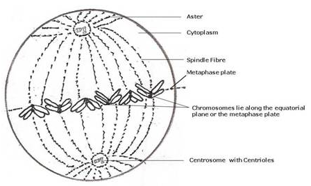
(d) Metaphase. The centromeres of chromosomes are drawn to the equator by equal pull of two chromosomal spindle fibres that connects each centromere to the opposite poles, forming a metaphasic plate.
(e)
| Mitosis | Meiosis |
| (i) Two daughter cells are produced. | (i) Four daughter cells are produced. |
| (ii) It is equational division i.e. the number of chromosome in the daughter cells or parent cells remains the same. | (ii) It is reductional division i.e. the number of chromosomes is reduced to half in the daughter cells. |
Solution D.5.
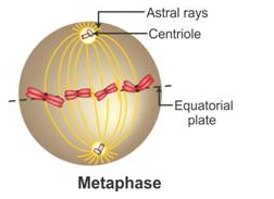
(a) Metaphase.
(b) 4.
(c) A – Animal
B – Animal
C – Plant
(d) (iv)
Solution D.6.
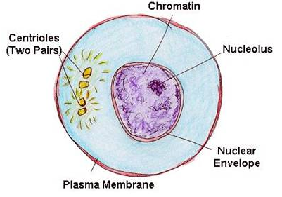
(a) This is an animal cell because:
- The outline is circular (in plants it would be angular {rectangular or polygonal}) and cell wall is absent.
- Centrosomes on centrioles are present. (These are found only in animal cells)
(b) Mitosis.
(c) B, C, D, A.
(d) Interphase.
(e)
More Resources for Selina Concise Class 10 ICSE Solutions